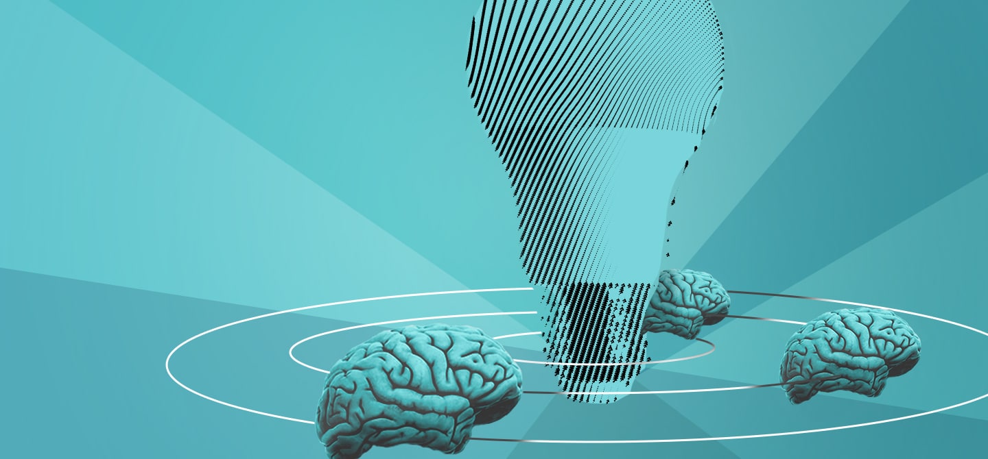

Are our brains hardwired at birth? At least, partially, says science.
Over the past few years, studies in the brain region responsible for memory, the hippocampus, suggests that the neurones there seem to be pre-wired during development 1. This leads to the conclusions that groups of neurones determined by very early events may form the elementary building blocks of our memory. To understand how these building blocks emerge during development, new advances in microscopy offer a way to see into the brain, allowing researchers to watch how the hippocampus changes as new experiences occur.
Painting neurones
Back in 2007, a team based at Harvard University developed a technique that allows researchers to colour neurones in a developing brain – which they aptly dubbed brainbow. As a result, when scientists look at them under a microscope, they see the brain as a striking collection of multi-coloured neural cells. Other than creating images that seem more like art than science, the technique allows researchers to point out specific neurones, precisely locating the trajectories of their spindly axons.

Using colour labels in this way enables researchers to probe neurogenesis, the process whereby the brain produces new neurones. As such, each cell is essentially a clone of a parent cell so, when labelled using brainbow, each will carry the same colour as their ancestor, providing a way to follow the lineage of brain cells.
Where there is HOPE…
If colouring neural cells is the first step, the second is being able to see them in high resolution. After all, the brain is composed of dense bundles of intertwined neurones – not so easy to pick out the detail. In a new European project, HOPE, set to take place from next year, Emmanuel Beaurepaire and his team at Institut Polytechnique de Paris are using three-photon microscopy to peer deep into the hippocampus.
Previous work from Pr. Rosa Cossart at Aix-Marseille University, a collaborator of HOPE, describes the process of pre-wiring of the hippocampus and the changes that come when new memories occur. The project will rely on three-photon microscopy to look at coloured neurones, specifically following changes in the hippocampus from birth in mice.
The team, which also includes Jean Livet from Institut de la Vision, plans to produce extensive maps of neuronal projections and study hippocampal structure to see how the cells connect with one another: a project which will require both human- and computer-power using deep learning.
“We want to see the way brain circuits are put in place during development and how that can be linked to experience in the adult brain,” says Emmanuel Beaurepaire. “Of course, as always with the brain, we are restricted by our ability to look inside it. Using three-photon microscopy – which we’ve been working on for the past four years – we can look about a millimetre deep into tissue of a live mouse.”
Powerful lasers are used to stimulate the fluorescent materials, meaning the colourful neurones can be seen with high precision. Not only can the technique look deeper than other microscopy methods, but its high sensitivity also allows researchers to see detail to the point of individual synapses. It might not seem like much, but 1 mm is certainly enough to take a good look around. “Our multi-photon microscopy is ideal for the hippocampus – of mice. The technique is too invasive for a larger brain like a human and, for now, we wouldn’t be able to go deep enough,” he states.
Going beyond the visual
“Neuroscience is the main application of multiphoton microscopy, but it will go hand-in-hand with other techniques,” he adds. “Further down the line, we also plan to use optogenetics, a technique which lets us stimulate or block specific neurons using light to observe the effects.”
“At the moment we are seeing a huge leap forward in our understanding of neuroscience – in terms of memory, consciousness and learning – which will no doubt lead to new solutions in a range of domains,” says Emmanuel Beaurepaire.
Problems that can occur in the developing hippocampus are known to be involved in a range of health problems that can persist throughout life: epilepsy and autism, to name a few. But going back to the original questions at the beginning of this article, the answer is one of fundamental science. “It is about first understanding how the brain works. Later, perhaps, they can be built upon for medical or psychiatric treatments.”