
Covid, pregnancy, heart attack: how to improve tests

- Lateral flow tests (LFA) are faster, cheaper, and less demanding than clinical laboratory tests.
- But they only provide a qualitative response such as “yes” or “no”, where a quantitative response would be valuable as well.
- The use of luminescent nanoparticles in LFA tests could improve their sensitivity by 40-50 times, while reducing their cost.
- This method is being developed with the aim of providing easy access to test results in any location and at any time.
- A small device has been developed to illuminate nanoparticles, facilitating the reading of the result obtained.
Since the Covid-19 pandemic, we have all become familiar with lateral flow tests (LFA), commonly referred to as “antigen tests”. This type of test can, in principle, detect the presence of any protein antigen in body fluids (urine, blood, saliva etc.) – not only SARS-CoV‑2. Their advantage over conventional tests performed in medical laboratories (such as ELISA) is that they are fast (15 minutes instead of several hours), inexpensive and do not require heavy laboratory equipment.
They are therefore useful for detecting many diseases, but also changes in the body such as pregnancy, or the presence of a protein food that could cause food poisoning. However, unlike ELISAs, one of the limitations of LFA tests is that they only give a qualitative answer. For a pregnancy test, for example, the results are still limited to “yes/no” – either you are pregnant or you are not. However, there are many cases where it would be useful to determine the amount of the protein being tested for. For example: “yes” you are pregnant, but for how many weeks? Or, “yes” you have Covid, but with what viral load?
Our method can be considered 40 to 50 times more sensitive than traditional ones.
Another limitation of LFA tests is that they are not particularly sensitive. Developing quantitative and sensitive LFA tests would push the boundaries of what these tests can be used for. They could even indicate how long ago a pregnancy started. This sensitivity and accuracy would also allow the test to detect the amount of cTnI protein naturally secreted before cardiac arrest, for example. Coupled with its new accessibility, it could be performed directly in the ambulance, facilitating patient management.
120x less antibody, 40x more sensitive
Fanny Mousseau, a researcher at the Optics and Biosciences Laboratory at Institut Polytechnique de Paris, is working on the development of LFA tests based on a new technology. LFA tests are based on the use of detection antibodies that attach to the targeted proteins. “The antibody (~10 nm) in this type of test must be coupled to something perceptible to the naked eye,” explains the researcher, “something larger and coloured, hence the use of gold nanoparticles (~50–100 nm) in commercial tests. The first step in our work was therefore to find a substitute with a more intense and quantifiable optical signal.”

The innovation: replacing gold nanoparticles with luminescent nanoparticles. “By using luminescent nanoparticles, we were able to improve the sensitivity of the test and make it quantitative and more robust.” And after some research into optimising the use of these nanoparticles, the Laboratory’s team managed to use 4x fewer of them than gold nanoparticles, which amounts to using 120x less detection antibody and thus considerably reducing the cost of a test. “When analysing LFAs made with our nanoparticles with the naked eye, the lowest detectable protein concentration is 5x to 10x lower,” says the researcher. “Using our optical analysis method, made with the help of the application we developed for this purpose, we can detect concentrations that are 2–4 times lower. With the two combined, our method can be considered 40–50 times more sensitive than the traditional one.”
In addition to this increase in sensitivity, using luminescent nanoparticles allows this test to become quantitative, instead of qualitative as with gold nanoparticles. “A test is considered accurate if after three repetitions we get the same measurement three times,” says Fanny Mousseau. “However, by accurately determining the quantity of targeted proteins present in the body, we can, for example, determine the stage of a disease and follow its evolution.”
Two tests in one, multiplexing
The Laboratory’s team is also working on the potential that this method has with regard to multiplexing – the possibility of detecting several proteins in a single test, and therefore in a single procedure. “In the case of certain pathologies, it is not the evolution of a single protein that is interesting, but that of several,” explains the researcher. For example, endocan is a biomarker of inflammation. This protein exists in two forms, ‘native’ and ‘cleaved’, and knowing the respective quantities of these two forms may make it possible to better define the treatment of certain pulmonary diseases. To decide on the most suitable treatment for the patient according to his other conditions, it is necessary to observe the quantity of “native” endocan and that of “transformed” endocan.
We are able to determine the presence of three different proteins in one multiplexed test with an accuracy rate of 30%.
Developing this potential is still a work in progress: at the moment the research team does not consider this method to be reliable enough for multiplexing. The reasons for this lack of reliability are known, however, and are due to the phenomenon of cross-reactivity. “For the detection of two proteins, two separate detection systems are needed [one for the antibody, another for the nanoparticles], which gives rise to the possibility of so-called cross-reactivity between the different systems.”
“So far, multiplexed tests are still qualitative. This year, I developed an innovative calibration curve, which takes this cross-reactivity into account, to try to read the results of a multiplex more accurately. As a result, we can determine the presence of three different proteins in one multiplexed test with an accuracy rate of 30%. This is a promising start,” she says. The researcher is optimistic that clinical trials will begin soon.
Virtually automated analysis
Once the technique for improving the sensitivity of LFA test strips had been identified, all the researchers had to do was develop an analysis method for these tests. “The main application of our research was to replace blood sampling,” says Fanny Mousseau. With our method, the results are quicker, but just as accurate. This method was developed with a view to facilitating these analyses in places where access to blood tests is limited [in war zones, in developing countries, etc.], or when they are urgently needed [in an ambulance in case of a heart attack, for example].
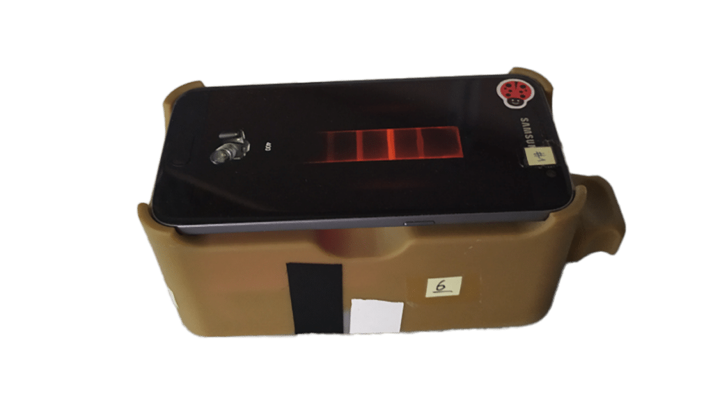
As a result, the ongoing research required a tool to illuminate the nanoparticles to make it easier to read the results. “The analysis methods required the use of huge equipment, which was contradictory to the portable advantage of this type of test,” explains the researcher. “In our laboratory, we invented a small device capable of performing the test. This small device, which is thicker but not larger than a mobile phone (~10 cm x 5 cm x 5 cm), comes with an application with which the user photographs the test.”
“We have integrated the analysis program. The user simply takes the picture and presses the control strip on the LFA strip (the strip that verifies that the test has worked). The algorithm, knowing the exact distance between the control strip and the test strip (the strip on which our protein of interest is detected) will analyse, pixel by pixel, the colour intensity of the test strip.
The result is then displayed, simply indicating the amount of target protein present in the test. “In the long term, the goal is to simplify the process as much as possible so that everyone can have access to it,” she concludes.