Contrary to what we may think, it is not only biological science research that contributes to advances in cancer therapy. Physics, and in particular particle science, has also contributed to important therapeutic advances; particularly involved in both improvements in effectiveness and reductions in side effects of radiotherapy, which accounts for more than half of all treatments.
Eliminating tumours
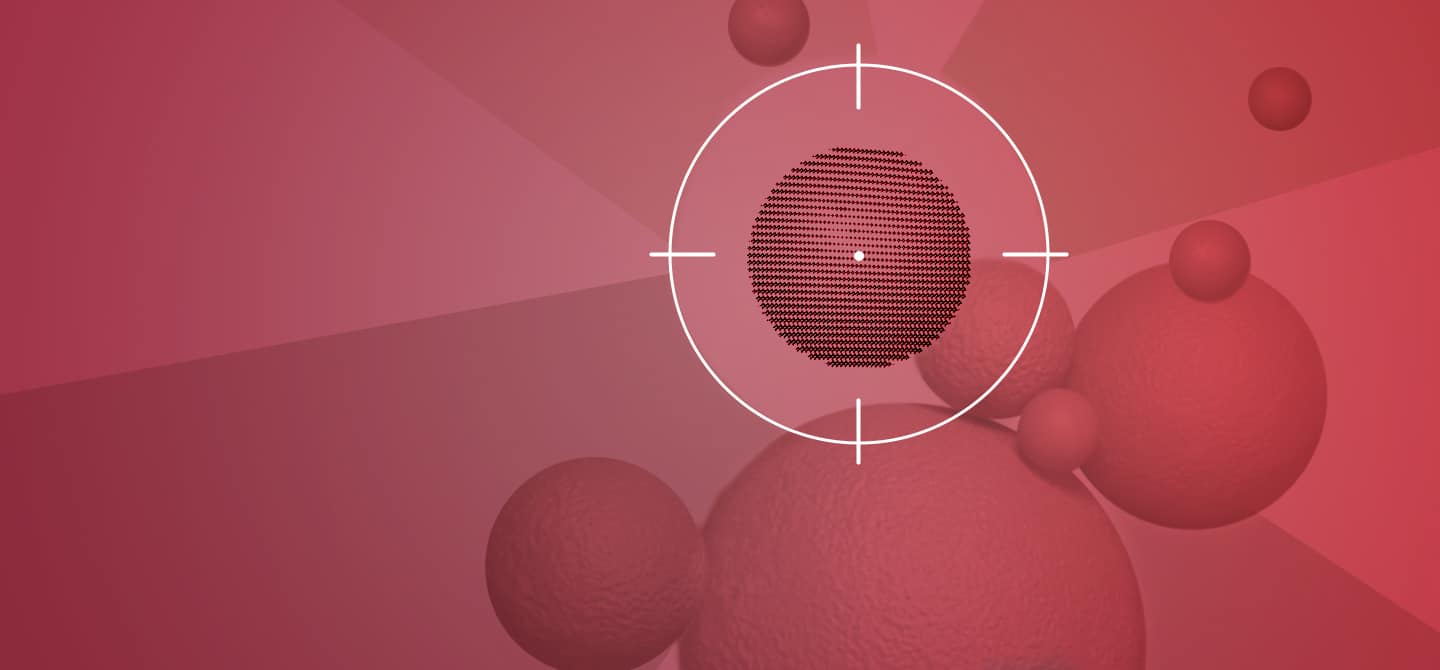
To understand this progress, we must first understand the principle of radiotherapy; an approach that consists of eliminating cancer cells using radiation. This radiation destroys the cancer cells as precisely as possible, without affecting the surrounding healthy cells. To achieve this, doctors rely on radiation-induced toxicity, i.e. the lower resistance of cancer cells to the effects of radiation compared to normal cells. This property ensures the therapeutic margin, the difference between an effective dose* and a toxic dose. The latter is also the result of topological advances and better beam focusing. The combination of these two phenomena, radiation-induced toxicity and topology, protects the healthy tissue around the tumour.
Ionising rays deposit energy deep in the tissue. They act at different levels: atomic, molecular, chemical, biological, and physiological. At the atomic level, radiation interacts with the chemical components contained within cells. Their ionising actions produce reactive species, such as free radicals, which can also destroy DNA and drive the cell to death. They also act directly at the molecular level, producing breaks in the DNA molecules. If there is enough damage, it overwhelms the cell’s self-repair processes. So, when the cell attempts to divide, as cancer cells do, it fails to complete its division and dies. This amount of damage attacks the structure of the tumour.
Several types of particles can be used: X‑rays (photons) are the most common. Since the 1990s, doctors have been using proton therapies, and more recently, the use of electron beams in cancer treatment has been studied.
Finding the right dose
The mechanisms of how radiotherapy works are known, and its biological control seemed clear. And it was thought that the biological effect was induced by the dose of radiation administered, a dose-response effect typical of biology. But recently this certainty has been overturned as it was discovered that the time profile of the dose alters the toxicity of the radiation1. This is known as the ‘flash effect’, which consists of delivering the dose of radiation in an extremely short time – over a few milliseconds instead of several minutes.
The shorter the time, the lower the sensitivity of healthy cells to the radiation, while that of cancer cells remains the same. The Flash effect thus increases the therapeutic margin. In practice, this reduces the undesirable effects of radiotherapy by causing less burning and producing less fibrosis – abnormal scarring of healthy tissue that can hinder the functioning of an organ, such as the liver or lungs when they are close to the irradiated area.
This effect is increasingly documented by clinical research, but the precise explanation is still missing, opening up a new field of research.
New lasers
This discovery has prompted scientists to re-explore sources of particles. In this respect, my team and I are studying the value of laser radiation. Unlike conventional systems, lasers are pulsed rather than continuous sources of particles. Laser sources have a different time profile to conventional or even Flash sources. The radiation generated is both very fast and very locally intense. We are currently trying to study its biological effect and it does not seem to be comparable to the Flash effect.
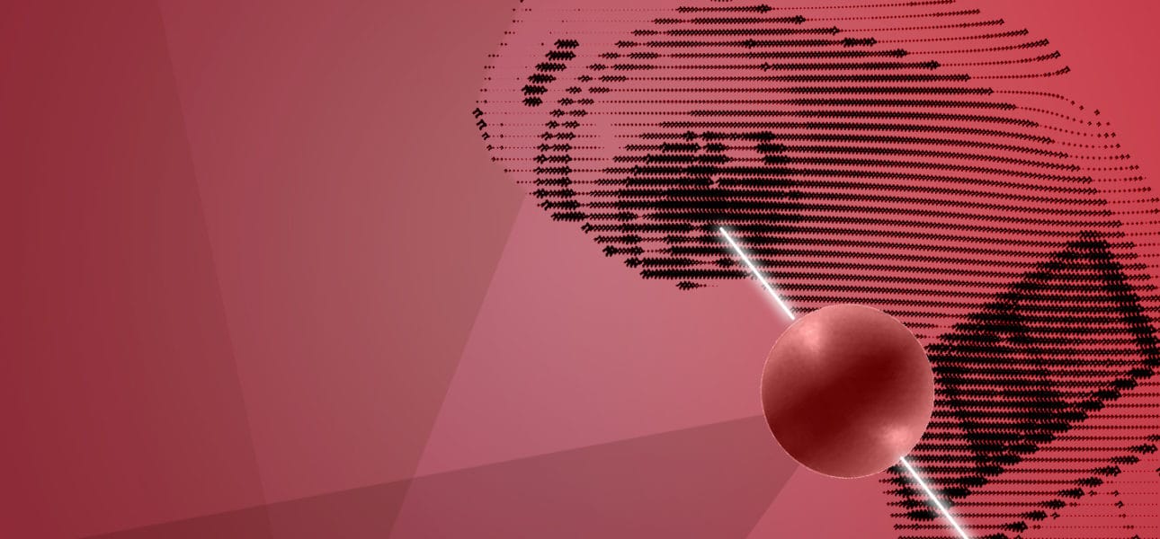
On cells in culture, we have observed a biological effect, a toxicity of cancer cells which seems interesting2. We are currently studying the feasibility of carrying out in vivo tests to better confirm this effect. Other progress is linked to advances in physics. This is the case with nanoparticles, which aim to concentrate irradiation locally. The idea is to inject these nanoparticles into the tumour. They potentiate the radiation and thus make it possible to administer a lower level of irradiation for a constant biological effect. This reduces the side effects, too.
Other nanoparticles release drugs when irradiated. They form an inert cage that traps cytotoxic molecules. Under the local action of radiation, the cage opens and releases the anti-cancer treatment. This approach is designed to prevent the patient from being subjected to general drug toxicity. Only the irradiated area is in contact with the cytotoxic molecules.
And that’s just to mention therapeutic progress because diagnosis, with the major progress made in cancer imaging, is another area that is fed by progress in the physical sciences.
* dose: the energy deposited on a mass of tissue