Layer by layer: how 3D printing is transforming hospitals and medical research
- 3D printing is increasingly being used in surgery, for example in the development of anatomical models tailored to each patient, enabling surgeons to rehearse operations in advance.
- In hospitals, the majority of 3D projects focus on obsolete hospital equipment, resulting in savings of between €120,000 and €140,000 per year.
- Since February 2023, Brest Hospital has developed its own 3D printing platform, W.Print, joining hospitals in Lyon, Besançon and Paris.
- Since 2021, the GI Jaw programme has been working on replacing missing bone in cleft lip and palate cases using a 3D-printed biomaterial tailored to patients’ anatomy.
- A recruitment programme for dogs with cleft lip and palate has been set up to develop a new 3D-printed surgical reconstruction technique.
Practising an operation before surgery, educating patients, adapting equipment to a disability, repairing obsolete technology… 3D printing has made its way into hospitals, benefiting patients, healthcare professionals and researchers alike, promising more personalised care and sustainable techniques.
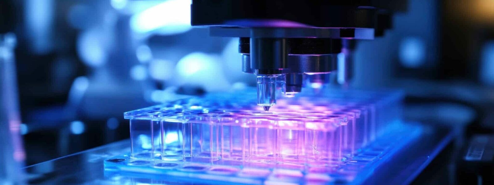
3D printing in surgery
Since February 2023, Brest Hospital has developed its own 3D printing platform, W.Print, joining hospitals in Lyon, Besançon and Paris. This new service builds on a research project originally led by Samuel Guigo on the management of complex intracranial aneurysms. “These operations require significant preparation beforehand. I therefore developed patient-specific 3D anatomical models, enabling surgeons to rehearse the operation, anticipate difficulties and adapt surgical techniques,” explains the researcher.
These models also help prepare surgical strategies. For example, in the case of a large tumour located beneath the eye – in the maxillary sinus – 3D printing makes it possible to plan the bone reconstruction required after removal, well in advance of the operation. More generally, healthcare professionals can thus practise on realistic anatomical models, sometimes with the addition of circuits simulating blood flow, or flexible or rigid materials to replicate the human body as closely as possible.
Currently the platform’s coordinator, Samuel Guigo oversees and supports nearly 80 projects a year. At this stage, we are not talking about bioprinting (where biological materials are generated for grafts, for example), or medical devices (which may be in prolonged contact with the body). We are simply talking about filament or resin printers that can create all sorts of objects. So why all the hype?
Training, explaining, repairing: 3D printing beyond the operating theatre
In addition to medical applications, the Brest-based platform has diversified to cover every aspect of hospital life, starting with patients and their families. When it comes to explaining a condition, a deformity or a surgical procedure, nothing beats a physical object. In cases of multiple disabilities in children, for example, these models become therapeutic education tools, helping families to better understand and engage with the care pathway. The platform is also part of the Rehab Lab network, dedicated to creating personalised assistive devices with and for people with disabilities. The aim: more sustainable, less expensive and better-utilised bespoke solutions, thereby facilitating a return home after hospitalisation.
3D printing helps tackle the problem of obsolete hospital equipment, with an estimated saving of between 120,000 and 140,000 euros per year
“But the majority of projects actually focus on the practical, day-to-day needs of the hospital,” reveals the coordinator. Pipette holders, adapted stands, spare parts unavailable on the market: 3D printing helps tackle the problem of obsolete hospital equipment. This results in estimated savings of between €120,000 and €140,000 per year, at a time when public hospitals are under significant budgetary pressure.
This approach is also driving a shift in working practices. Care assistants, stretcher bearers, catering staff and administrative personnel now come forward with their specific needs to co-design solutions. For Samuel Guigo, this is a way of reducing the “irritants” of daily life, those minor issues that undermine the working atmosphere, and of fostering a culture of innovation among all staff. Today, requests from other hospitals are flooding in, and Samuel Guigo, who is currently the only person managing the entire initiative, is sharing his expertise with other institutions whilst training students in 3D design.
Research: a shift in perspective
In Nantes, an ongoing multidisciplinary research project provides a concrete illustration of the potential and challenges of 3D printing in healthcare. Bringing together Nantes University Hospital, the Oniris VetAgroBio Veterinary School and Inserm’s RMeS laboratory, the GI Jaw programme has been focusing on the treatment of cleft lips and palates since 2021.
Today, these congenital malformations, characterised by a partial or complete absence of bone between the mouth and the nose, are mainly treated using bone grafts taken from the child’s hip. This is an effective solution, but one that nevertheless involves post-operative pain and an additional scar for young patients who undergo surgery as early as five or six years of age. “We need 2 or 3 ml of bone, so sometimes we have to supplement it with bone substitute,” adds Pierre Corre, professor of maxillofacial surgery at Nantes University Hospital and one of the three project leaders. “That’s where the clinician’s challenge becomes the researcher’s!” says Baptiste Charbonnier, a research engineer at Inserm 1, the second member of the trio.
The idea behind the project is simple: to replace the missing bone without taking it from elsewhere, using a 3D-printed biomaterial perfectly adapted to each patient’s anatomy. Why not use ceramic or the standard bone cement used in dental procedures? “Because this malformation of the upper jaw is located in a complex area, at risk of infection between the mouth and the nose: the aim is to create perfect contact so that the blood vessels of the native bone can rapidly colonise the implant and enable deep bone regrowth,” explains Pierre Corre. A new material therefore needs to be developed that is malleable during implantation, hardens once in place and is eventually resorbed to make way for new bone.
The aim is to create perfect contact so that the blood vessels from the native bone can rapidly colonise the [3D-printed] implant and enable deep bone regrowth
For this study (and to complete the trio), Pierre Maitre, a veterinarian and senior lecturer in surgery at the Oniris Veterinary School, has initiated a recruitment drive for dogs with cleft lip and palate, which are usually euthanised at birth due to this malformation, to develop a new surgical reconstruction technique using 3D printing. For this study – and to complete the trio – Pierre Maitre, a vet and senior lecturer in surgery at the Oniris Veterinary School, has organised a recruitment drive for dogs with cleft lip and palate, which are usually euthanised at birth due to this malformation, to develop a new surgical reconstruction technique using 3D printing.
It all begins with a CT scan: the implant is digitally designed to precisely match the bone defect and the surrounding mucosa, and to facilitate insertion during surgery. “After printing, the cemented implant dries and is then sterilised,” explains Baptiste Charbonnier. To make it malleable, the biomaterial is rehydrated in the operating theatre. This hydration also allows it to be impregnated with the patient’s bone marrow, which will become the new colonising cells, and triggers the setting of the cement, which will allow the implant to harden after insertion. Once in place, the 3D implant then becomes a temporary scaffold, gradually colonised by the patient’s bone cells.
The results are promising – after six months, the implant is almost entirely resorbed, replaced by natural bone. Trials have already been conducted on 18 dogs, which could become ambassadors for this new technique among future young patients. But the researchers remain realistic: “We still have a higher failure rate than with autografts, but we have observed that bone regeneration appears to be greater with this new material, so that is already progress!”
The project, funded by the ANR for a further four years, is now entering a phase of material refinement and regulatory approval, with the aim of becoming a (biodegradable) Class 3 medical device. Every year in France, around 1 in 700 children is born with a cleft lip and palate, the majority of whom have a bone defect. Whilst the technique is not yet ready for routine clinical use, it already points the way towards more personalised, less invasive and less painful treatment for the young patients of tomorrow.